
Key Takeaways
Pictures can help you notice and document a change, but they cannot diagnose cancer. Many harmless growths resemble malignant tumors, while some serious cancers develop inside the body and leave little visible evidence. A veterinarian should assess any new, changing, bleeding, painful, or persistent abnormality.
- A photo shows appearance, not the nature of a growth.
- Benign and malignant tumors can look remarkably alike.
- Changes in size, color, texture, and behavior are worth recording.
- Skin findings may need aspiration, biopsy, or staging tests.
- Prompt veterinary attention is safest when warning signs appear.
What dog cancer pictures can—and cannot—tell you
Searching for types of dog cancer pictures can help a worried owner compare broad visual patterns, but online images are not a diagnostic tool. Lighting, fur, camera angle, and the dog’s skin color can all change how a lesion appears. More importantly, similar-looking growths may have very different causes. Use pictures to notice change and prepare useful information for a veterinarian, not to decide what a mass is at home.
Why appearance alone cannot confirm cancer
A photograph can show whether a lesion is raised, discolored, open, or surrounded by hair loss. It cannot show the cells beneath the surface or reveal whether a mass has invaded nearby tissue. Even an experienced person may mistake an irritated cyst, wart, abscess, or healing injury for a tumor. Appearance is only one clue, and it needs to be interpreted alongside the dog’s history and physical examination.
How benign growths can resemble malignant tumors
Harmless lumps may be smooth, firm, soft, pigmented, or inflamed after a dog licks them. Some grow slowly for years; others become suddenly swollen because of friction or infection. Cancerous tumors can also be small and quiet at first, so size alone does not settle the question. The visual distinction between benign and malignant growths is often too subtle for a reliable home judgment.
When a photo is useful for tracking changes
A dated photograph can make gradual change easier to recognize than memory can. Take the image from the same angle, in similar light, and include a ruler or familiar object nearby without pressing on the growth. Record where it is located, how long it has been present, and whether your dog is licking or guarding it. A simple timeline can help the clinic understand whether the change was sudden or gradual.
Why veterinary examination and testing matter
A veterinarian can feel the mass, examine nearby skin and lymph nodes, and decide whether testing is appropriate. Fine-needle aspiration, biopsy, or other investigations may identify cells and guide treatment. The Merck Veterinary Manual’s skin tumor overview also explains why tumor type, location, size, and the dog’s overall health affect management. A picture starts a conversation; it does not replace the examination.
Common types of skin cancer in dogs
Skin tumors in dogs do not share one standard appearance. Some sit on the surface, while others begin deeper in the tissue and only later change the skin above them. The examples below describe patterns a person might encounter in educational photographs, not a checklist for self-diagnosis.

Mast cell tumors and their variable appearance
Mast cell tumors are known for a wide range of appearances. They may look like a smooth lump, a raised and reddened area, or a mass that changes size after irritation. Swelling, itching, or repeated licking can make the surface look more dramatic than it did initially. A veterinary guide to mast cell tumors in dogs can provide context, but only sampling can determine what a particular lump contains.
Squamous cell carcinoma on the skin
Squamous cell carcinoma may appear as a crusted, scaly, ulcerated, or nonhealing area, sometimes with little obvious lump. It is more concerning when a sore repeatedly reopens or fails to behave like an ordinary minor injury. Areas with sparse hair or less pigment may receive more sun exposure, though a veterinarian must assess the individual lesion. This guide to sun-related dog skin cancer includes visual discussion of persistent sores and squamous cell carcinoma.
Melanoma in the mouth, skin, and nail bed
Melanoma involves pigment-producing cells and may appear dark brown, black, gray, or unevenly colored, but not every melanoma is deeply pigmented. In the mouth, a lesion may be difficult to see until there is bleeding, bad breath, drooling, or trouble eating. A nail-bed lesion can cause swelling or changes around one toe. Color is a useful observation, not proof of tumor type.
Soft tissue sarcomas beneath or on the skin
Soft tissue sarcomas often begin below the skin as a firm or rubbery mass. The overlying skin may look normal, especially early on, and the edges may be difficult to judge by touch. Some are slow-growing and some change more noticeably. Their depth and relationship to surrounding structures are reasons a veterinarian may recommend imaging or tissue sampling rather than relying on a photograph.
Basal cell tumors and other surface growths
Basal cell tumors and other surface growths can be rounded, raised, hairless, or attached by a narrow base. Many are benign, but an unusual appearance does not establish that they are harmless. A growth that catches on a collar, bleeds after scratching, or changes in texture deserves an examination. Educational dog tumor stock photos can illustrate variety, but stock imagery should never be used to match a diagnosis.
How different dog cancers may look in pictures
Pictures tend to draw attention to what is easiest to see: a lump, a wound, a patch of color, or a swollen body part. Those features can be helpful when describing a problem, especially if several images show progression. Still, a visible pattern can occur with many conditions, including infection, trauma, allergies, and benign growths. The safest approach is to describe what changed without assigning a diagnosis.
Lumps that are raised, flat, firm, or soft
A lump may be dome-shaped, flat against the skin, soft beneath the fingers, or unusually firm. It may move freely or seem fixed, although owners should not repeatedly squeeze or manipulate it. Note its approximate size and position instead. A mass that is new or changing should be assessed even when the dog seems comfortable.
Ulcers, bleeding, crusting, and nonhealing sores
An ulcer may look wet, raw, scabbed, or surrounded by irritated skin. Bleeding can result from a tumor, but it can also follow scratching, licking, or minor trauma. Repeated discharge and a sore that does not heal normally are more useful warning signs than a single photograph. Keep the area clean and prevent persistent licking while arranging veterinary advice.
Dark, pigmented, or irregularly colored lesions
A dark spot may be normal pigmentation, a freckle, dried blood, or a growth. Irregular borders, rapid enlargement, several shades within one lesion, or a new location can make it worth checking. Photographing the area in natural, even light helps avoid shadows that exaggerate color differences. Do not scrape away a crust or apply human skin products unless a veterinarian advises it.
Swelling around the mouth, eyes, paws, or nail beds
Swelling in these locations can affect eating, vision, walking, or nail growth, so even a small lesion may matter. A mass near the eye should not be pressed, and a mouth lesion may be difficult to inspect safely at home. Paw or nail-bed changes can also reflect infection or injury. Location, discomfort, and loss of function should guide how quickly you seek care.
Hair loss and skin changes around a growth
Hair may thin or fall out over a lump because the skin is stretched, inflamed, or repeatedly licked. Redness, thickening, scaling, or a change in temperature can add useful context. These signs are not specific to cancer, and allergic or infectious skin disease can look similar. The skin tumor information from the veterinary manual is a helpful reminder that diagnosis depends on more than surface appearance.
Cancer types that may not be visible on the skin
Not every cancer produces a photograph-friendly lesion. Some tumors develop in bone, glands, lymph nodes, or abdominal organs, where early changes may be felt only by a veterinarian or noticed through behavior. A dog may also have nonspecific symptoms rather than a visible mass. That is why ongoing changes in appetite, weight, energy, breathing, or movement deserve attention even when the skin looks normal.
Bone cancer and limb swelling
Bone cancer can be associated with persistent lameness, pain, or swelling in a limb. The visible change may be subtle at first, and a photograph cannot show what is happening inside the bone. Sudden severe lameness also has many possible causes, including injury. A veterinarian may recommend radiographs or other tests when pain persists or returns.
Mammary tumors along the belly
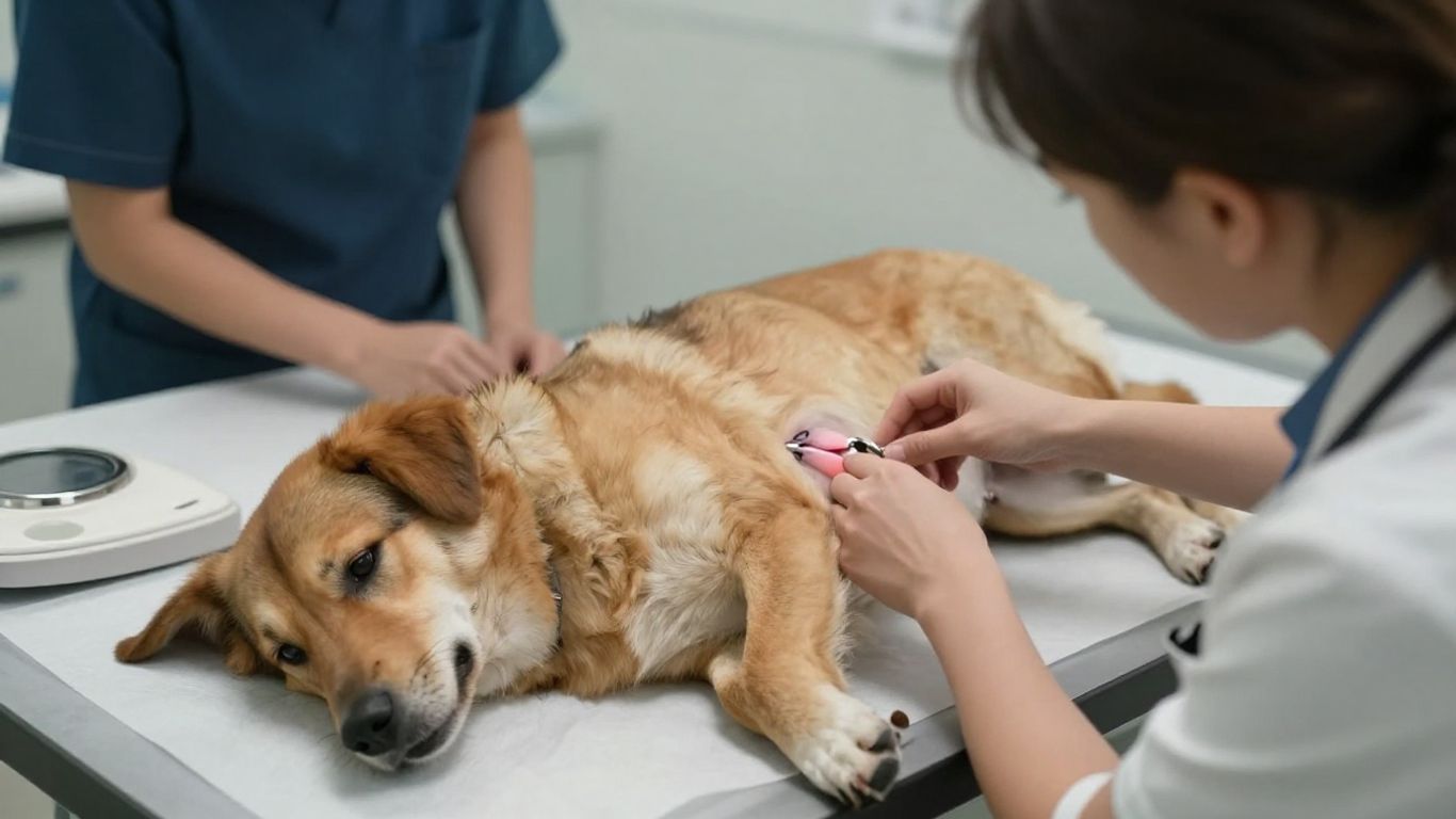
Mammary tumors may feel like one or more nodules along the chain of tissue on the underside of the dog. Some are easy to see; others are only detectable by gently examining the area. Avoid repeated pressing, which can irritate the tissue and make accurate assessment harder. New mammary lumps should be discussed with a veterinarian, particularly if they enlarge, ulcerate, or become painful.
Lymphoma and enlarged lymph nodes
Enlarged lymph nodes can occur under the jaw, in front of the shoulders, behind the knees, or in other locations. They may not be visible at all, but a veterinarian may detect them during a routine examination. Lymph-node enlargement can have infectious and inflammatory causes as well as cancer. If several nodes enlarge or the dog also seems unwell, prompt evaluation is sensible.
Abdominal tumors and changes in body shape
Tumors inside the abdomen may cause a fuller-looking belly, discomfort, vomiting, reduced appetite, or changes in stool. A change in body shape can also reflect weight gain, fluid, pregnancy, or another medical problem. Do not press firmly on a distended abdomen. A clinic may use blood tests and imaging to find the source safely.
Lung or internal cancers with nonspecific symptoms
Coughing, reduced stamina, faster breathing, or unexplained tiredness can come from many conditions, including heart and airway disease. Internal cancer is only one possibility, and a photograph of the dog’s exterior is unlikely to clarify the cause. Persistent or worsening symptoms need a proper examination. For a broader reminder of subtle changes owners may miss, see these signs of cancer in dogs.
Warning signs that need prompt veterinary attention
A single unusual mark does not automatically mean an emergency, but certain changes should not be watched indefinitely. Speed, recurrence, pain, and effects on normal function are particularly useful clues. When in doubt, call the veterinary clinic and describe what you see. A clear photo, measurement, and timeline can help staff advise you about urgency.
A lump that grows or changes quickly
Rapid enlargement, a sudden change in firmness, or a new irregular outline deserves prompt attention. The same is true when a previously quiet lump becomes red, warm, itchy, or painful. Do not wait for it to become very large before arranging an appointment. Growth rate is information a veterinarian can use when deciding what to test.
Repeated bleeding, discharge, or ulceration
A mass that repeatedly bleeds, produces fluid, or develops an open sore should be examined. Covering it tightly or applying household remedies may trap moisture and worsen irritation. If bleeding is heavy or does not stop with gentle pressure, contact an emergency clinic. Otherwise, protect the area from licking and arrange timely veterinary care.
Pain, limping, licking, or sensitivity
Behavior often reveals a problem before a visual change becomes obvious. Reluctance to climb, limping, repeated licking, flinching, or guarding a body part can indicate pain. These signs are not specific to cancer, but they should not be dismissed because the skin lesion looks small. A painful growth or persistent limb change warrants professional assessment.
Appetite, weight, energy, or bathroom changes
Unexplained weight loss, reduced appetite, unusual tiredness, vomiting, diarrhea, constipation, or altered urination can accompany many illnesses. Keep notes about frequency and duration rather than relying on memory. Four useful observations to bring to the appointment are:
- when the change began;
- whether it is worsening or intermittent;
- what your dog is eating and drinking;
- whether sleep, exercise, stool, or urine has changed.
These details help the veterinarian place a skin finding in the context of the whole dog. They can also reveal that a seemingly isolated lump is occurring alongside a broader health change.
Breathing difficulty, vomiting, or persistent coughing
Breathing difficulty is urgent, especially if the gums look pale, gray, or blue, the dog collapses, or breathing remains labored at rest. Persistent coughing and repeated vomiting also need veterinary attention, even though they have many causes unrelated to cancer. Seek emergency help when symptoms are severe, sudden, or accompanied by collapse. Do not delay care while trying to identify a matching picture online.
What happens after you find a suspicious growth
Finding a growth can feel frightening, but the next step is usually a methodical examination rather than an immediate assumption. Gather the dates, photos, measurements, and symptoms you have noticed. Avoid squeezing, cutting, or attempting to drain the area. The veterinarian will decide which information and tests are most appropriate for your dog.
How veterinarians examine and measure a mass
The appointment may include a full physical examination, careful palpation, measurement in more than one direction, and inspection of nearby lymph nodes. The veterinarian may ask when the growth first appeared and whether it has changed after rubbing or licking. Location, depth, mobility, and the condition of the overlying skin all matter. These observations help determine whether sampling or imaging should come next.
When fine-needle aspiration or biopsy is recommended
Fine-needle aspiration uses a small needle to collect cells for examination and may be suitable for many accessible masses. A biopsy removes a tissue sample and can provide more detailed information when aspiration is inconclusive or when the mass needs a more precise assessment. The choice depends on the suspected problem, location, size, and the dog’s comfort. Ask what the test can answer and what its limitations are.
Why staging tests may be needed
If cancer is identified or strongly suspected, staging tests may look for involvement elsewhere in the body. Depending on the case, these may include blood work, imaging, lymph-node sampling, or other evaluations. Staging is not required for every lump, and the veterinarian will tailor the plan to the likely tumor and the dog’s overall condition. The results help clarify treatment choices and expectations.
Questions to ask about diagnosis and treatment
It is reasonable to ask for plain-language explanations and written instructions. Useful questions include what the sample showed, whether margins or spread are a concern, what alternatives exist, and how quickly a decision is needed. Also ask about pain control, wound care, follow-up, and signs that should prompt a call. A second opinion may be appropriate if the diagnosis or options remain unclear.
How to photograph and monitor a growth safely
Take a wide image showing the body location and a closer image showing texture and color. Use consistent lighting, keep the camera parallel to the skin, and place a ruler beside—not on—the growth. Do not delay care while collecting a perfect series of pictures. A short educational video can also help explain how veterinarians assess suspicious masses.
After photographing the area, note the date, measurement, symptoms, and any change in behavior. Avoid irritating the growth with repeated handling, and follow the clinic’s advice about preventing licking. A calm record is more useful than frequent checking that makes the dog uncomfortable.
For owners balancing several household health concerns, the practical value of consistent routines appears in resources such as diabetic dog meal timing, though diabetes care is separate from evaluating a tumor. The same principle of recording changes applies to other decisions too, from planning a home landscape to preparing a villa project, but neither home project guidance nor photographs can replace veterinary testing. Likewise, improving a stagnant home listing and checking a roof for hail damage are unrelated tasks; they are included here only as examples of why careful observation should lead to the right professional.
Conclusion
Dog cancer pictures can help you recognize that a change deserves attention and create a useful record for the clinic, but they cannot tell you whether a growth is benign or malignant. New, changing, painful, bleeding, or persistent abnormalities should be assessed by a veterinarian, especially when appetite, energy, breathing, movement, or bathroom habits also change. Early, calm action gives you clearer information and more choices.
Frequently Asked Questions
Can a picture confirm that my dog has cancer?
No. A picture can document appearance and change, but diagnosis usually requires a veterinary examination and sometimes cell or tissue testing.
What does dog skin cancer usually look like?
It may appear as a lump, ulcer, crusted sore, discolored patch, or swelling, but these appearances also occur with noncancerous conditions and injuries.
Should every lump on a dog be tested?
Not every lump needs the same test, but a veterinarian should assess new or changing growths and decide whether monitoring, aspiration, biopsy, or another test is appropriate.
Is a fast-growing lump always cancer?
No. Infection, inflammation, irritation, and other problems can enlarge quickly. Rapid growth still warrants prompt veterinary evaluation.
Can internal cancers be present without a skin lump?
Yes. Some cancers affect bone, lymph nodes, abdominal organs, lungs, or other tissues and may first cause nonspecific changes such as tiredness, weight loss, pain, or altered breathing.
How should I photograph a suspicious growth?
Use even lighting, take a wide and close view, include a ruler for scale, and record the date. Do not press, scrape, or irritate the growth to improve the photograph.
When should I seek emergency care?
Seek urgent help for breathing difficulty, collapse, uncontrolled bleeding, severe pain, repeated vomiting, pale or blue gums, or any sudden severe deterioration.





Leave a Reply