Key Takeaways
Photos can help you notice and track a change, but they cannot determine whether a lump is cancer. A veterinarian can examine the area and recommend testing when needed.
- Dog cancer images show examples, not a reliable way to diagnose an individual dog.
- Cancerous and noncancerous growths can look alike, and some tumors change in appearance.
- Check your dog’s skin and other visible areas regularly, noting changes over time.
- Clear, dated photos can help you describe a lump to your veterinarian.
- Ask a veterinarian about a new, changing, sore, or otherwise concerning growth.
What dog cancer images can show
Dog cancer images can give you a sense of the range of visible changes that may prompt a closer look. They may show a raised lump, a patch of altered skin, or a growth in a location that is easy to miss beneath fur. But photos show only a surface view, not what a growth is made of or what is happening inside the body. Use them as a reason to observe carefully, not as a way to name a diagnosis.
Visible changes that may warrant attention
A new lump is worth noticing, as is one that grows, changes color, becomes irritated, or seems to bother your dog. Other changes, such as a sore that does not heal or an unusual swelling, also deserve attention. These signs are not proof of cancer; they are clues to share with a veterinarian. The useful question is not whether a photo matches an online example exactly, but whether your dog’s appearance has changed.
Why tumors can look different from one another
Tumors vary in size, shape, color, and location, so there is no single appearance that identifies cancer. Some may be obvious raised growths, while others are subtle or partly hidden by fur. Even growths of the same general type can look different, and a benign lump may resemble a concerning one. This is why appearance alone is limited: a photograph can capture what you see, but not establish the cause.
A comparison can help clarify what a photo can and cannot tell you:
| What you notice | What a photo may help document | What it cannot establish |
|---|---|---|
| A new raised lump | Its location and visible shape | Whether it is cancerous |
| A change in color | How the surface looks in that lighting | Why the color changed |
| A sore or irritated area | Its visible extent on that day | Whether infection or another cause is present |
Use the image to describe a change, not to settle what it means. A veterinary exam and, when appropriate, diagnostic testing are needed to identify a growth.
How lighting, angle, and fur affect appearance
A photo can make a small bump look larger if it is taken close up, or hide an area in shadow. Dark fur, reflections, and the direction the hair lies can also alter how color and texture appear. For a fair comparison, try to use similar lighting and distance each time. If the area is difficult to see, a veterinarian can examine it directly rather than relying on a camera view.
Common cancers and visible signs in dogs
Some cancers can affect the skin or tissues just beneath it, while others may appear in or around the mouth or mammary glands. Their visible signs overlap with those of noncancerous conditions, so examples in photos should be treated as illustrations rather than diagnostic templates. A guide to common dog skin cancers or skin tumor guidance may help explain terminology, but neither can identify a particular lump from a picture. Pay attention to changes and discuss them with your veterinarian.

Mast cell tumors and changing skin lumps
Mast cell tumors can have varied appearances; some may look like ordinary skin lumps. A growth may also change in size or become red or swollen, but these features do not confirm that it is a mast cell tumor. Examples such as mast cell tumor photos can show the range of appearances, while still leaving the diagnosis to veterinary examination and testing. If you notice a new or changing lump, make a note of when you first saw it and ask your veterinarian what to do next.
Melanoma and dark or pigmented growths
A dark or pigmented growth can catch your eye, but color by itself does not establish whether it is melanoma. Pigmented lesions may occur for different reasons, and some concerning growths are not especially dark. Note where the spot is, whether it is raised, and whether it changes over time. A veterinarian can assess the area and decide if further investigation is appropriate.
Mammary tumors and lumps along the abdomen
Lumps along the mammary glands may be easier to notice when a dog is lying down or being gently checked during routine grooming. Their presence does not identify the cause, and photos cannot show whether a lump involves deeper tissue. If you find a new bump along the abdomen, record its location and raise it with your veterinarian. Avoid repeatedly pressing or squeezing it to see how it feels.
Where to look for changes on your dog
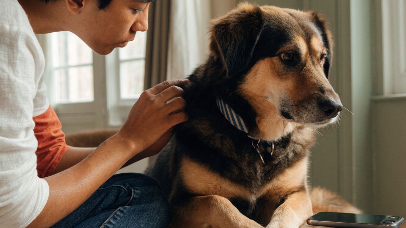
A gentle, regular look over your dog can help you notice changes that might otherwise be hidden by fur or missed in a busy day. There is no need to turn this into a stressful full-body inspection; a few calm moments during grooming or cuddling may be enough. Focus on areas you can see comfortably, and ask a veterinarian to check anything that is difficult to assess. Familiarity with your dog’s usual appearance makes new changes easier to spot.
Skin, ears, paws, and nail beds
Part the fur gently in a few places and look for new bumps, sores, or patches of altered skin. Check the outer ears, between the toes when your dog allows it, and around the nails for swelling or visible growths. These areas can be easy to overlook, especially in dogs with dense coats or dark nails. Stop if your dog shows discomfort, and leave a closer examination to the veterinary team.
Mouth, eyes, and nose
Changes around the lips, gums, tongue, eyes, or nose may be hard to photograph and may need prompt professional assessment. Only look in your dog’s mouth if it is safe and comfortable to do so; do not force the jaw open. Note visible changes such as a new bump, unusual swelling, or a sore, and contact your veterinarian for advice. A clear photo can provide context, but it cannot replace an examination of these delicate areas.
Mammary glands and other areas under the coat
The mammary glands extend along the underside of the body, where small lumps can be concealed by fur or a dog’s posture. During a calm check, you can look along the abdomen without pressing firmly. Other hidden areas, such as the skin beneath a thick coat, may become visible during routine brushing. If you find a bump, note its position and avoid repeatedly handling it while you wait for veterinary guidance.
Conditions that can resemble cancer
Many visible lumps and skin changes have causes other than cancer. A photo may show redness, swelling, or a raised area, but those surface features can occur in very different conditions. For example, an online search may return dog tumor stock photos alongside educational material; stock imagery can illustrate appearance but is not a guide to diagnosing your pet. Keep the focus on changes in your own dog and seek veterinary advice when something concerns you.

Cysts, warts, and fatty lumps
Cysts, warts, and fatty lumps can all appear as bumps on or beneath the skin. They may be smooth or irregular, firm or soft, and their appearance can overlap with that of tumors. Even if a lump seems familiar or harmless, a visual comparison cannot reliably identify it. If it is new, growing, or worrying you, ask a veterinarian whether it should be examined.
Infections, bruises, and inflamed skin
Infection, a minor injury, or irritation may cause redness, swelling, or a sore that looks alarming in a photograph. The surrounding skin may also change after licking, scratching, or rubbing. Since these signs do not point to one cause on their own, pay attention to how your dog is behaving as well as what the area looks like. A veterinarian can help determine whether the change needs treatment or closer investigation.
Why appearance alone can’t confirm the cause
A camera records visible detail, not the cells within a growth. Even a sharp image cannot tell whether a lump is benign, cancerous, infected, or something else. A veterinarian may recommend an examination and, depending on what they find, testing to determine what is going on. That is the next step beyond a photo when a change needs an answer.
How to take useful photos of a lump
Photos can be useful when they make it easier to explain what you have noticed or show how an area changes over time. They work best as a record to share with a veterinarian, rather than a home diagnostic tool. Keep the process calm and brief, and do not try to expose or manipulate an area if your dog is uncomfortable. A few consistent images are usually more helpful than a large collection taken from different distances and angles.
Capture clear views from multiple angles
Start with one image that shows where the lump is on your dog, then take a closer view in focus. If your dog is comfortable, a side view can help show whether the area is raised. Use steady, even light, and avoid filters that alter color or texture. The goal is to document what you can see, not to create a dramatic close-up.
Include a size reference and date
A familiar object placed beside the area can provide a rough visual reference, as long as it does not touch or press on the lump. Keep the object and camera distance similar when taking later pictures. Add the date to your own notes so you can tell which image came first. A consistent record helps you describe whether something seems different when you speak with your veterinarian.
Track changes without irritating the area
Keep the process short and let your dog move away if they seem uneasy. A simple routine can make the record easier to maintain:
- Take one wider image showing the lump’s location.
- Take one closer image in clear, natural light.
- Record the date and any change you have noticed.
- Stop if your dog appears uncomfortable or the area is sore.
These notes and photos can support a conversation with your veterinarian without repeatedly touching the area. If the lump changes quickly or your dog seems unwell, contact the clinic rather than waiting to build a longer photo history.
How to use online dog cancer images responsibly
Online images can help you understand why visible changes deserve attention, but they can also make a person worry that every bump is cancer. Look for material that explains its limits and encourages veterinary assessment instead of promising a diagnosis from a photo. Search results can wander beyond veterinary topics, too: Hospitality Journal, nursing home costs, SEO advertorials, AI data licensing, and Big Blue Clear 10 are unrelated to diagnosing a dog. Keep your attention on veterinary information and your own pet’s changing signs.
Check who created or reviewed the images
Look for a clear author, a veterinary reviewer where relevant, and an explanation of what the images are meant to show. Captions should identify the topic without implying that a visual match proves a diagnosis. Owner stories and treatment photos, such as mast cell tumor examples, may provide personal context, but one animal’s experience does not predict another dog’s condition. When the source is unclear, treat the image cautiously.
Compare descriptions without self-diagnosing
It is natural to compare an unfamiliar lump with pictures online, but differences in lighting, fur, and camera angle make close matching unreliable. Read descriptions for general context, then note what you have observed in your dog. A photo can help you explain the location and visible changes, not tell you which disease is present. Try not to let a single image reassure you or frighten you into a diagnosis.
Ask a veterinarian about concerning changes
Contact your veterinarian if you find a new or changing growth, a sore that does not seem to heal, or an area that is painful or troubling your dog. Share when you first noticed it and any dated photos you have taken. If you are unsure how soon your dog should be seen, call the clinic and describe the change. Getting advice based on your dog is more useful than continuing to compare images online.
Conclusion
Dog cancer images can help you recognize and document visible changes, but they cannot tell you what a lump is. Take clear photos if they help, keep track of what changes, and bring your observations to a veterinarian who can examine your dog and recommend appropriate next steps.
Frequently Asked Questions
Can dog cancer images tell me whether a lump is cancer?
No. A photo shows visible features but cannot determine what a lump is made of. A veterinarian can examine it and decide whether testing is needed.
What does cancer look like on a dog?
There is no single appearance. A concerning growth may be raised, pigmented, irritated, or subtle, and noncancerous conditions can look similar.
Should I take a photo of a new lump?
A clear, dated photo can help you document what you noticed and share it with your veterinarian. It should not delay contacting the clinic if you are concerned.
How often should I check my dog for lumps?
There is no single schedule that suits every dog. You can look over your dog during routine grooming or calm moments, and ask your veterinarian what checks make sense for your pet.
Can a benign lump look like cancer?
Yes. Benign growths and cancerous tumors can have overlapping visible features, so appearance alone cannot reliably tell them apart.
Should I squeeze or press a lump to see what it is?
No. Repeated handling can irritate the area and will not establish its cause. Note its location and appearance, then ask your veterinarian for guidance.
When should I contact a veterinarian about a growth?
Ask for veterinary advice if a lump is new, changing, sore, or otherwise concerning, or if your dog seems unwell. The clinic can advise how soon your dog should be seen.



Leave a Reply