
Key Takeaways
Testicular changes in a dog should be assessed by a veterinarian, even when the dog seems comfortable. Early examination helps clarify whether a change is a tumor, inflammation, trauma, or another condition.
- Testicular tumors are most often found in older, intact male dogs.
- Retained or undescended testicles increase the risk of certain tumors.
- Swelling, firmness, hair loss, or mammary enlargement can be warning signs.
- Neutering is usually the main treatment, with tissue sent for analysis.
- Prognosis depends on tumor type, spread, hormone effects, and overall health.
Understanding testicular cancer in dogs
Testicular cancer in dogs refers to abnormal growth arising in one or both testicles. Some tumors remain localized and are found incidentally, while others produce hormones or cause visible changes. The word “cancer” can sound alarming, but the outlook varies considerably between tumor types and individual dogs.
Testicular tumors are primarily a concern in intact male dogs, particularly as they grow older. A veterinarian may recommend examination, imaging, and removal of the affected tissue to determine exactly what is present.
What testicular tumors are
A testicular tumor develops when cells in the testicle multiply abnormally. It may appear as a discrete mass, a general enlargement, or an area that feels different from the surrounding tissue. Some dogs have no obvious discomfort, so a seemingly minor change still deserves attention.
A mass cannot be classified reliably by touch alone. Infection, bruising, torsion, cysts, and other disorders may produce similar findings. Tissue examination after surgery is often needed for a definitive diagnosis.
The most common types of testicular tumors
Veterinarians commonly discuss seminomas, Sertoli cell tumors, and interstitial cell tumors, also called Leydig cell tumors. They arise from different cell populations and may behave differently. Some are associated with hormone production, while others mainly cause a change in the testicle itself.
A brief comparison can help explain why the pathology report matters:
| Tumor type | Cell origin or association | Possible clinical pattern |
|---|---|---|
| Seminoma | Germ cells | Often presents as a testicular mass |
| Sertoli cell tumor | Sertoli cells | May produce estrogen-related signs |
| Interstitial cell tumor | Leydig cells | May produce testosterone or remain clinically quiet |
| Mixed tumor | More than one cell population | Behavior depends on the components present |
These broad categories are not a substitute for an individual diagnosis. A veterinary pathologist evaluates the removed tissue and reports features that help guide follow-up.
For additional background, this canine tumor overview discusses the main tumor categories and the role of undescended testicles.
How age and retained testicles affect risk
Testicular tumors are more often identified in older, unneutered dogs than in young dogs. Risk is also higher when one or both testicles remain in the abdomen or groin rather than descending into the scrotum, a condition called cryptorchidism.
A retained testicle is exposed to a different internal environment and may be harder for an owner to examine. It can also make surgery more involved. Dogs with a retained testicle should have the issue discussed with a veterinarian rather than simply monitored indefinitely.
Whether testicular cancer spreads to other parts of the body
Many canine testicular tumors are treated successfully when they remain localized, but spread is possible in some cases. The likelihood depends on the tumor type, its cellular features, its size, and whether there is evidence of invasion or metastasis.
Possible areas of concern include nearby lymph nodes and organs in the abdomen or chest. This is why a veterinarian may recommend staging tests even when the visible change appears confined to one testicle. The clinical management literature offers a more detailed testicular tumor review for readers seeking a veterinary research perspective.
Recognizing the signs and symptoms
Signs of testicular cancer in dogs can be subtle or absent, especially early on. Owners may notice a difference while bathing, grooming, or giving medication, but many changes are first identified during a routine examination. A new finding should not be dismissed simply because the dog is eating, walking, and playing normally.
The appearance of symptoms can also depend on whether a tumor affects hormone production. Some dogs develop changes in the coat or mammary tissue, while others show only an enlarged or unusually firm testicle.

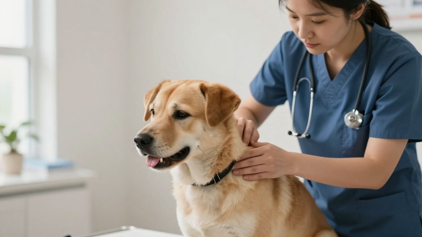
This photograph reflects the kind of calm, hands-on assessment that may reveal an abnormality before a dog appears seriously ill.
Changes in testicle size, shape, or firmness
One testicle may become larger, smaller, harder, or less symmetrical than the other. The surface may feel irregular, or the entire testicle may seem heavier. Comparing both sides gently can be useful, but owners should avoid repeated pressing if the area appears painful.
A sudden change can have causes other than cancer, including inflammation or trauma. Even so, any persistent difference warrants a veterinary examination.
Scrotal swelling, pain, or skin changes
Swelling of the scrotum may result from a mass, fluid, inflammation, bleeding, or twisting of the testicle. Redness, thickened skin, licking, or sensitivity can also signal a problem. Severe pain or rapid enlargement should be treated as urgent.
Dogs do not always vocalize when they hurt. Restlessness, reluctance to sit, frequent licking, or a change in gait may be the only clues that the area is uncomfortable.
Hormone-related signs such as hair loss or enlarged mammary glands
Some tumors alter the balance of reproductive hormones. Depending on the hormones involved, a dog may develop symmetrical hair loss, darker skin, a thinner coat, enlarged mammary glands, or changes in the penis or prepuce.
Behavioral changes, including increased interest in other dogs or irritability, can occur but are not specific. Hormone-related signs should be considered alongside the physical findings rather than used to identify a tumor on their own.
General symptoms that may indicate advanced disease
A dog with more extensive illness may lose weight, tire easily, eat less, vomit, or show breathing difficulty. Enlarged lymph nodes, abdominal discomfort, or weakness can have many causes, so these signs do not prove that a testicular tumor has spread.
Still, prompt veterinary assessment matters when general illness appears alongside a testicular change. Earlier evaluation gives the care team more options for confirming the diagnosis and planning treatment.
How veterinarians diagnose the condition
Diagnosis begins with a careful examination and a history of what the owner has observed. The veterinarian considers the dog’s age, reproductive status, previous injuries, medication history, and the timing of any changes. The goal is not only to find a mass but also to understand the dog’s overall condition.
Testing is selected according to the examination findings. Some dogs need only a focused workup before surgery, while others require additional evaluation to look for hormone effects or possible spread.
Physical examination and medical history
The veterinarian palpates the scrotum, both testicles, the groin, and nearby lymph nodes. If a testicle is retained, the groin and abdomen may also be examined for its location. The clinician may look for mammary enlargement, coat changes, prostate-related signs, or other hormone effects.
An owner’s observations can be surprisingly helpful. Note when the change began, whether it is growing, whether the dog licks the area, and whether appetite, energy, urination, or behavior has changed.
Blood tests and hormone evaluations
Routine blood work can help assess general health before anesthesia and surgery. It may also reveal anemia, changes associated with illness, or other findings that affect treatment planning. In selected cases, hormone testing helps investigate signs such as hair loss or mammary enlargement.
Blood tests rarely identify the tumor type by themselves. Their main value is placing the physical finding in context and making anesthesia or further testing safer.
Ultrasound and other imaging tests
Ultrasound can show the internal structure of the testicle and may help distinguish a focal mass from more diffuse changes. It can also be useful when a testicle is retained in the abdomen or groin. Chest radiographs or abdominal imaging may be recommended when the veterinarian needs to assess possible spread.
Imaging findings guide decisions, but they do not always provide a final answer. A suspicious area may still require removal and microscopic examination.
Biopsy, tissue analysis, and staging
In many cases, the affected testicle is removed and submitted to a laboratory. The pathologist identifies the tumor type, examines its cellular behavior, and may comment on features associated with recurrence or spread. Staging combines this information with the examination and imaging results.
A pathology report is therefore more than a label. It helps the veterinarian explain the outlook, decide whether additional treatment is warranted, and design an appropriate monitoring schedule. A separate pathology discussion describes the major histological groups in more technical detail.
Treatment options for affected dogs
Treatment depends on the tumor’s location, the dog’s health, whether a testicle is retained, and whether disease has spread. For a localized tumor, surgery is commonly both diagnostic and therapeutic. The veterinary team will discuss anesthesia, pain relief, and the likely extent of the procedure beforehand.
Owners may understandably focus on removing the visible mass, but the surrounding findings matter too. A complete plan accounts for the other testicle, possible abdominal surgery, and any hormone-related complications.

The procedure and aftercare should be tailored to the dog rather than based on the appearance of the mass alone.
Why neutering is usually the primary treatment
Removal of both testicles, commonly called neutering or castration, is usually the primary treatment for canine testicular tumors. It removes the known tumor and prevents disease from developing in the remaining testicle. It also eliminates the main source of reproductive hormones.
The removed tissue is generally sent for histopathology. Even when surgery appears straightforward, the laboratory result helps determine whether further staging or monitoring is needed.
When surgery may require additional planning
A retained abdominal testicle may require an abdominal approach rather than a routine scrotal procedure. A very large mass, suspected vascular involvement, or other health problems may also change the surgical plan. Pre-operative blood work and imaging help the veterinarian anticipate these issues.
The surgeon may discuss referral to a specialty hospital when the anatomy is complicated or when advanced imaging and intensive monitoring are appropriate. The safest plan is the one matched to the dog’s findings and anesthetic risk.
Treatment for tumors that have spread
If staging suggests metastasis, treatment may involve referral to a veterinary oncologist. Options can include additional surgery, chemotherapy, radiation in selected situations, or careful monitoring, depending on the tumor’s biology and location.
There is no single protocol that suits every dog with advanced disease. The care team weighs potential benefit against side effects, travel, cost, and the dog’s comfort and quality of life.
Managing pain, infection, and other complications
Pain control is planned around the operation and adjusted as the dog recovers. Antibiotics may be used when infection is present or when the veterinarian believes they are indicated, but they are not automatically needed for every uncomplicated procedure.
Hormone-related problems may require separate attention, particularly if they have affected the skin, bone marrow, prostate, or mammary tissue. Owners should ask which signs are expected and which ones should prompt a call.
Recovery and aftercare
Most dogs need a quiet recovery period after neutering, even if they seem eager to resume normal activity. The incision may look mildly pink or swollen at first, but it should be checked every day. Following the discharge instructions closely helps protect the surgical site and makes complications easier to spot.
Recovery is also a time to observe the dog’s appetite, comfort, urination, and energy. A gradual return to normal behavior is reassuring; a sudden decline is not.
What to expect after surgery
Sleepiness during the first day can follow anesthesia and pain medication. Mild reluctance to eat may occur, although many dogs resume eating relatively quickly. The veterinarian will explain when water, food, medication, and normal toileting should resume.
The surgical area should gradually become less swollen and less sensitive. A follow-up visit may be scheduled to assess healing and review the pathology report.
Incision care and activity restrictions
Keep the incision dry and prevent licking, chewing, running, and rough play while the tissues heal. A protective collar or recovery garment may be needed. Short, controlled leash walks are usually safer than unrestricted activity during the early period.
A simple routine can help owners stay consistent:
- Check the incision at least once daily.
- Give prescribed medication exactly as directed.
- Use leash walks for bathroom breaks.
- Keep bedding clean and the recovery area calm.
These measures reduce disruption to the incision and make it easier to notice a change. If the dog is unusually uncomfortable despite medication, contact the veterinary practice for advice rather than adding a human pain reliever.
Follow-up examinations and monitoring
The first recheck focuses on wound healing, comfort, appetite, and activity. Later appointments may be based on the pathology report and the risk of recurrence or metastasis. Some dogs need periodic physical examinations and imaging, while others require less intensive follow-up.
Bring the pathology report and any discharge instructions to future veterinary visits. Keeping a short record of weight, energy, appetite, and new physical changes can make monitoring more useful.
When post-operative symptoms need urgent attention
Call a veterinarian promptly if the incision opens, bleeds continuously, becomes increasingly hot or swollen, produces discharge, or develops a bad odor. Repeated vomiting, collapse, severe pain, pale gums, difficulty breathing, or inability to urinate also require urgent attention.
Do not wait for a scheduled recheck when symptoms are rapidly worsening. The surgical team would rather assess a concerning change early than have an owner worry at home.
Prognosis and factors that affect outcomes
The outlook for a dog with a testicular tumor cannot be determined from the mass alone. Tumor type, grade, size, location, hormone production, and evidence of spread all contribute. General health and the ability to recover from surgery matter as well.
Many localized tumors have a favorable outcome after surgical removal, but every pathology report tells a slightly different story. The veterinarian can translate the report into a practical follow-up plan.
How tumor type influences the outlook
Seminomas, Sertoli cell tumors, and interstitial cell tumors do not behave identically. Some remain localized, while others are more likely to cause hormone-related disease or show concerning microscopic features. Mixed tumors may require especially careful interpretation because more than one cell population is involved.
The report may also describe whether the tumor was completely removed and whether its appearance suggests aggressive behavior. Those details are more useful than relying on the tumor name alone.
The role of early detection and staging
Finding a tumor before it causes widespread illness generally gives the veterinary team more treatment choices. Staging helps identify whether nearby lymph nodes, the lungs, or abdominal organs need closer evaluation.
Early detection does not guarantee a particular outcome, but it can prevent a localized problem from going unnoticed. This clinical management resource provides further general context on diagnosis, treatment, and prognosis.
Signs that may suggest a poorer prognosis
A large or rapidly growing mass, marked systemic illness, evidence of metastasis, or aggressive features on histopathology may make the outlook more guarded. Persistent hormone-related complications can also affect recovery and quality of life.
These factors are not predictions on their own. A dog with one concerning feature may still respond well, while a quiet-looking mass may require more investigation than expected.
Long-term monitoring after treatment
Monitoring usually includes physical examinations and review of any new testicular, skin, mammary, respiratory, or appetite changes. The schedule is individualized according to pathology and staging. Owners should keep follow-up appointments even when the dog appears completely recovered.
If a new lump, enlarged lymph node, unexplained weight loss, or persistent cough develops, report it rather than waiting for the next routine visit. Ongoing observation is practical, not a reason to live in fear.
Reducing risk and protecting your dog’s health
Owners cannot prevent every testicular tumor, but they can reduce avoidable risk and improve the chance of early discovery. Knowing whether both testicles descended, scheduling routine examinations, and discussing neutering are sensible parts of preventive care.
Health decisions should reflect the dog’s age, breed, medical history, lifestyle, and reproductive role. A veterinarian can explain timing and alternatives in the context of that individual dog.
The importance of checking for retained testicles
Both testicles should normally be identifiable in the scrotum. If one is absent, unusually small, or located in the groin or abdomen, ask a veterinarian to confirm its position. A retained testicle is not simply a cosmetic variation; it carries increased health concerns and may be difficult to monitor at home.
Do not attempt to locate or manipulate a suspected retained testicle yourself. Imaging or surgery may be needed to determine where it is and how best to manage it.
Routine veterinary examinations
Regular examinations allow a veterinarian to compare findings over time and identify changes that owners may miss. The visit can include palpation of the testicles, lymph nodes, prostate region, skin, and abdomen, along with a discussion of behavior and general health.
Routine care is also a useful time to review parasite prevention, dental health, weight, and vaccination needs. A broad examination can place a testicular concern in the context of the dog’s whole health.
When to discuss neutering with your veterinarian
Neutering should be discussed as an individual decision, particularly for a dog with a retained testicle or a known testicular mass. The veterinarian can explain likely benefits, surgical approaches, timing, and any risks related to age or other medical conditions.
If a dog is intended for breeding, that conversation still matters. A suspected tumor or retained testicle should be evaluated before breeding plans continue.
Why new testicular changes should be evaluated promptly
A new lump, asymmetry, swelling, or change in firmness may be benign, but waiting to see whether it disappears can delay diagnosis. Contact the veterinary practice and describe what you noticed, when it began, and whether the dog seems painful or unwell.
A prompt appointment does not mean cancer is certain. It means the dog can receive the right examination while the finding is still easier to characterize and manage.
Conclusion
Testicular cancer in dogs can range from a quiet, localized tumor to a condition involving hormones or distant disease. Checking for changes, arranging timely veterinary care, and following through with pathology and monitoring give owners the clearest path forward. When in doubt, a new testicular abnormality is worth a professional examination.
Frequently Asked Questions
Is testicular cancer common in dogs?
Testicular tumors are among the more common tumors found in intact male dogs, especially older dogs. The exact risk varies with age, reproductive status, breed, and whether a testicle is retained.
Can a dog have a testicular tumor without showing symptoms?
Yes. Some dogs have no obvious pain or illness, and the change may be noticed only during grooming or a routine examination. Lack of symptoms does not rule out a tumor.
Does a retained testicle increase cancer risk?
Yes. An undescended or retained testicle has a higher risk of developing certain tumors than a testicle positioned normally in the scrotum. Discuss evaluation and management with a veterinarian.
How is a testicular tumor confirmed?
A veterinarian may use physical examination, blood work, ultrasound, and other imaging. In many cases, definitive classification comes from microscopic analysis of tissue removed during surgery.
Is surgery usually successful for testicular tumors?
Surgery is often effective when the tumor is localized and the dog is otherwise a suitable anesthetic candidate. The pathology report and staging results are needed to give a more individualized outlook.
Can testicular tumors spread in dogs?
Some can spread, although the likelihood depends on tumor type and other microscopic and clinical features. Imaging and laboratory analysis help determine whether spread is suspected.
When should I call a veterinarian about a testicular change?
Arrange an examination for any new enlargement, lump, firmness, asymmetry, swelling, pain, or skin change. Seek urgent care for severe pain, rapid swelling, collapse, breathing difficulty, or heavy bleeding.




Leave a Reply