Key Takeaways
Photos can help you notice and document a change, but they cannot determine whether a growth is cancerous. Many harmless lumps resemble tumors, so a veterinarian needs to examine anything new, changing, bleeding, or persistent. Careful photographs, measurements, and a clear timeline can make that appointment more useful.
- A picture is a starting point, not a diagnosis.
- Canine tumors vary widely in color, texture, size, and location.
- Benign lumps, infections, cysts, and allergies can look alarming.
- Veterinary testing is the only reliable way to identify a suspicious growth.
- Do not squeeze, cut, or medicate a lump at home.
Understanding what images of dog cancer can and cannot show
Searching for images of dog cancer may help you compare general patterns and recognize that concerning changes are worth taking seriously. Still, online photographs show only one moment, often under unfamiliar lighting and from an unknown angle. The safest use of an image is to guide observation and encourage a timely veterinary conversation, not to label a mass yourself.
Why photos are useful for awareness
A clear photograph creates a visual record that can be compared over days or weeks. It may help you notice that a spot has enlarged, changed color, opened, or begun affecting nearby skin. A visual gallery such as dog tumor images can also help owners understand how varied these growths may appear, although no gallery can replace an examination.
Photographs are especially useful when a dog has a heavily coated body or a small lesion that is easy to lose track of. Take several views, including one that shows the surrounding anatomy, so your veterinarian can understand the location rather than seeing an isolated close-up.
Why visual appearance cannot confirm cancer
The surface of a lump does not reveal its cell type, behavior, or depth. A smooth, movable mass may be benign, while a small or ordinary-looking bump may still deserve testing. Conversely, redness, swelling, or a crust can result from irritation rather than cancer.
Appearance alone is not diagnostic. A veterinarian may need to feel the growth, assess nearby lymph nodes, and collect cells or tissue before discussing what it is and how it should be managed.
Conditions that can resemble cancerous growths
Several noncancerous problems can look surprisingly similar in a photograph. Cysts, lipomas, warts, abscesses, insect reactions, allergic skin disease, and healing injuries may all create a raised, red, hairless, or crusted area. Even different types of tumors can overlap in appearance, which is why a visual comparison has limits.
For a broader look at the difference between common masses and more serious possibilities, readers sometimes consult common dog tumors. Use that kind of resource to prepare questions, not to make a final diagnosis from a screen.
When an image should prompt a veterinary appointment
Arrange an appointment when a growth is new, persists, changes, or worries you. A prompt visit is particularly sensible when the area is ulcerated, repeatedly traumatized, rapidly enlarging, or located where it interferes with walking, eating, breathing, or normal grooming.
The pattern matters more than a single snapshot. Keep the original images and note when you first saw the change, whether your dog reacts to touch, and whether the area has altered between photographs.
Common types of canine skin tumors and masses
Canine skin masses include both benign and malignant conditions, and their appearances can overlap. Some are firm and fixed; others are soft and mobile. Location, growth rate, skin changes, and the dog’s overall health all add context, but they still cannot establish a diagnosis without testing.

Mast cell tumors
Mast cell tumors are among the skin growths veterinarians commonly investigate. They may resemble a small, quiet lump or appear irritated, swollen, or inflamed. Their size and surface can fluctuate, so a photograph taken on one day may not capture the whole pattern.
A changing mass should not be dismissed because it sometimes looks smaller. Your veterinarian can decide whether sampling is appropriate and whether additional evaluation is needed.
Melanomas and pigmented growths
Melanomas and other pigmented growths may be dark, but not every dark bump is a melanoma, and some melanomas are not strongly pigmented. Growths around the mouth, lips, nail beds, or other sensitive areas deserve careful attention because they can affect everyday function.
Color is only one observation to record. Note texture, attachment to deeper tissue, bleeding, and whether your dog is rubbing or licking the area. A guide to dog skin cancer signs may help organize those observations before an appointment.
Soft tissue sarcomas
Soft tissue sarcomas can present as firm masses beneath or within the skin. They may be slow-growing and difficult to assess from the surface, particularly when a dog has thick fur. A seemingly stable lump still merits professional evaluation if it is new or continues to enlarge.
The veterinarian may measure the mass and consider how freely it moves over deeper structures. Those physical findings help determine which diagnostic steps are most useful.
Benign lumps such as lipomas and cysts
Lipomas are often soft, rounded, and movable, while cysts may have a more defined surface or contain material. These descriptions are common visual patterns, not guarantees. A benign-looking lump can become irritated, and a malignant mass can initially look unremarkable.
The practical question is not whether a lump resembles a familiar picture. It is whether the growth has been properly identified and whether its location or behavior creates a problem for the dog.
What dog cancer may look like on different body areas
A growth’s body location can change the signs an owner notices first. On a torso, the earliest clue may be a bump beneath the coat; on a paw, it may be persistent licking or a changed gait. Looking at the whole dog, rather than only the visible spot, provides a more useful picture.
Skin and coat changes
On the skin, a concerning area may appear as a raised nodule, a patch of hair loss, a persistent sore, a scab that returns, or a region of altered color. Some lesions are obvious, while others are hidden under fur and found only during brushing or routine handling.
Part the coat gently in good light and compare both sides of the body. Avoid repeatedly rubbing the spot, since handling can cause irritation and make its appearance harder to interpret.
Mouth, nose, and facial growths
Growths around the mouth or nose may cause bad breath, drooling, facial swelling, noisy breathing, sneezing, or difficulty picking up food. A mass inside the mouth may not be visible in an ordinary portrait, and dogs often resist close inspection when an area hurts.
Do not force the mouth open for a photograph. If there is bleeding, trouble breathing, or an inability to eat or drink, contact a veterinarian promptly rather than waiting for a better image.
Mammary glands and the abdomen
Lumps along the mammary chain or abdomen can be subtle at first. Run your hands gently over the area during a calm routine, noting any firm nodule, swelling, skin change, or discharge. Avoid pressing repeatedly, especially if the dog shows discomfort.
A simple record can make changes easier to communicate. The following observations are useful to bring to an appointment:
- The date the area was first noticed.
- Its approximate length and width.
- Whether it feels soft, firm, movable, or fixed.
- Changes in appetite, energy, grooming, or comfort.
That information does not diagnose the mass, but it gives the clinical examination a clearer timeline.
Paws, nails, and between the toes
A growth near a nail bed or between the toes may first look like a minor injury. Persistent licking, swelling, a split nail, discharge, or reluctance to bear weight can be more noticeable than the growth itself. Paw lesions are also easily irritated by walking and repeated licking.
Look briefly after outdoor activity and keep the area clean, but do not dig between the toes or apply human creams unless a veterinarian recommends them. A lesion that does not settle should be examined.
Warning signs to look for beyond a visible lump
Cancer can cause signs that are not limited to a bump on the skin, although these same signs can have many other explanations. A dog may seem uncomfortable, lose interest in food, or tire more easily because of pain, infection, dental disease, or another illness. Patterns, persistence, and change from normal are the most useful clues.

Changes in size, shape, or color
Measure a growth rather than relying on memory. Changes in diameter, height, outline, pigmentation, or firmness are worth recording, especially if they occur over a short period. A mass that repeatedly changes after being touched or irritated also deserves professional attention.
Use the same ruler, lighting, and camera distance when possible. Consistent records are more informative than a series of dramatically different close-ups.
Bleeding, ulceration, discharge, or odor
A surface that breaks open, bleeds easily, produces fluid, or develops an unpleasant odor may be inflamed, infected, traumatized, or affected by a tumor. These signs do not identify the cause, but they do mean the area should not be ignored.
Keep your dog from chewing or scratching the lesion if you can do so comfortably, and ask the veterinary team how to protect it. Do not place harsh disinfectants or household products on an open area.
Pain, itching, licking, or loss of function
Repeated licking, scratching, rubbing, limping, reluctance to jump, or guarding a body part can signal discomfort. A facial or oral growth may change eating, while a paw lesion may alter the dog’s stride. Some tumors are painless, so a lack of reaction does not make a growth harmless.
Watch how the symptom affects ordinary behavior. A short video of limping, coughing, or difficulty eating can sometimes provide useful context, as long as recording does not delay care.
Weight loss, fatigue, coughing, or appetite changes
Whole-body changes deserve attention even when no obvious lump is present. Unexplained weight loss, persistent fatigue, reduced appetite, vomiting, coughing, or breathing changes can result from numerous conditions, including some cancers. They are reasons to arrange a veterinary assessment rather than reasons to assume the worst.
For perspective, a skin-mass photo guide can help explain why visual patterns overlap among different conditions. It should be paired with an examination, particularly when local changes occur alongside a decline in general health.
How veterinarians diagnose suspicious growths
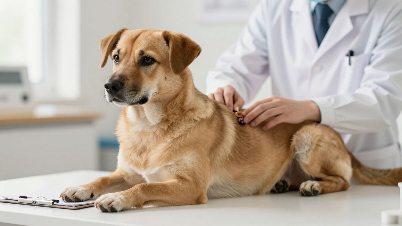
Diagnosis usually begins with a hands-on assessment and develops according to what the veterinarian finds. The process may be simple for a small, clearly characterized mass or more involved when a lesion is deep, fast-growing, or associated with other signs. Your veterinarian can explain the purpose, limits, and timing of each test.
Physical examination and medical history
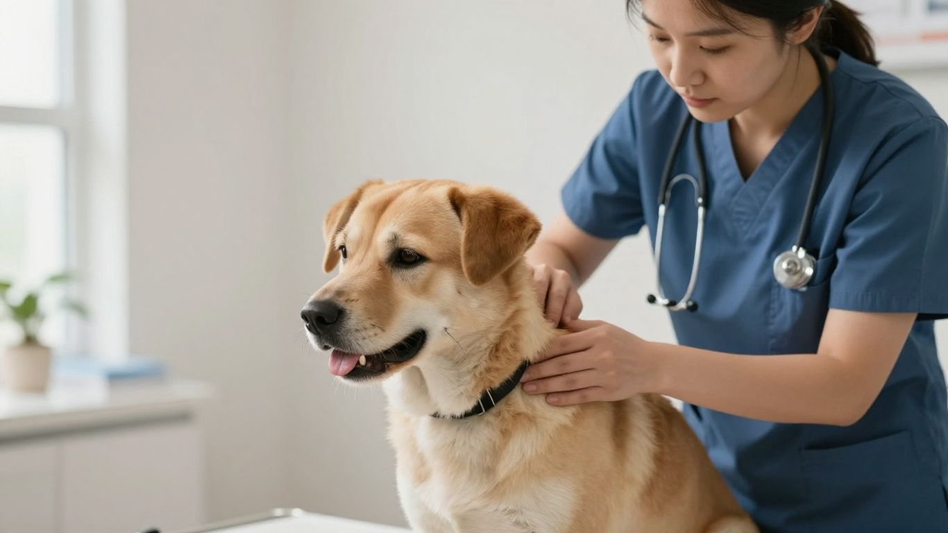
The veterinarian will consider when the growth appeared, how it has changed, whether it bothers the dog, and whether there are other symptoms. They may measure it, examine its surface, assess mobility, and check the rest of the skin and nearby lymph nodes.
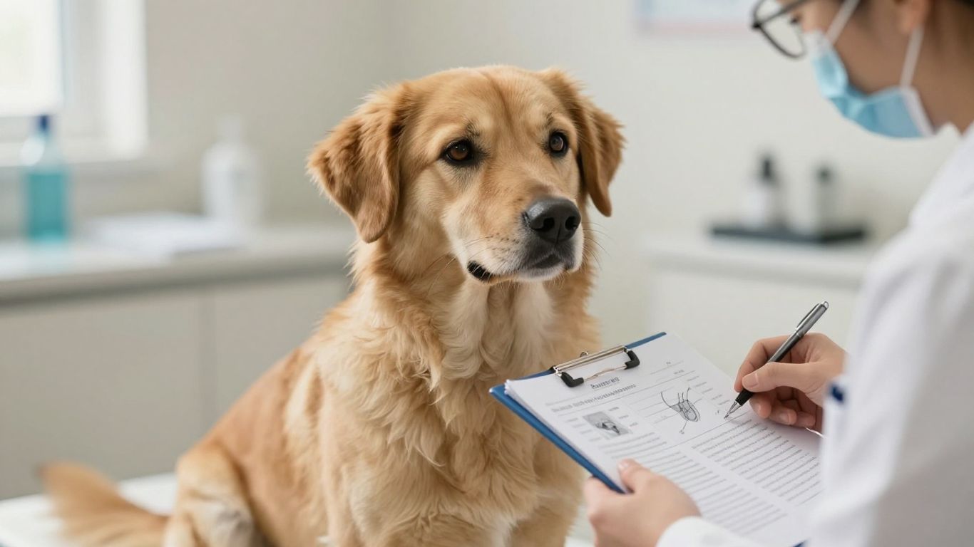
Bring photographs, measurements, medication details, and any notes about changes at home. A consistent history can help distinguish a recent irritation from a longer-standing problem.
Fine-needle aspiration and biopsy
A fine-needle aspiration collects cells from a mass for examination. Depending on the result and the clinical context, the veterinarian may recommend a biopsy, which evaluates a tissue sample in greater detail. Sometimes an initial sample is inconclusive and another method is needed.
The choice depends on the growth’s location, size, accessibility, and suspected behavior. Avoid assuming that a needle sample or biopsy is unnecessary simply because a mass looks ordinary in a photograph.
Blood tests and diagnostic imaging
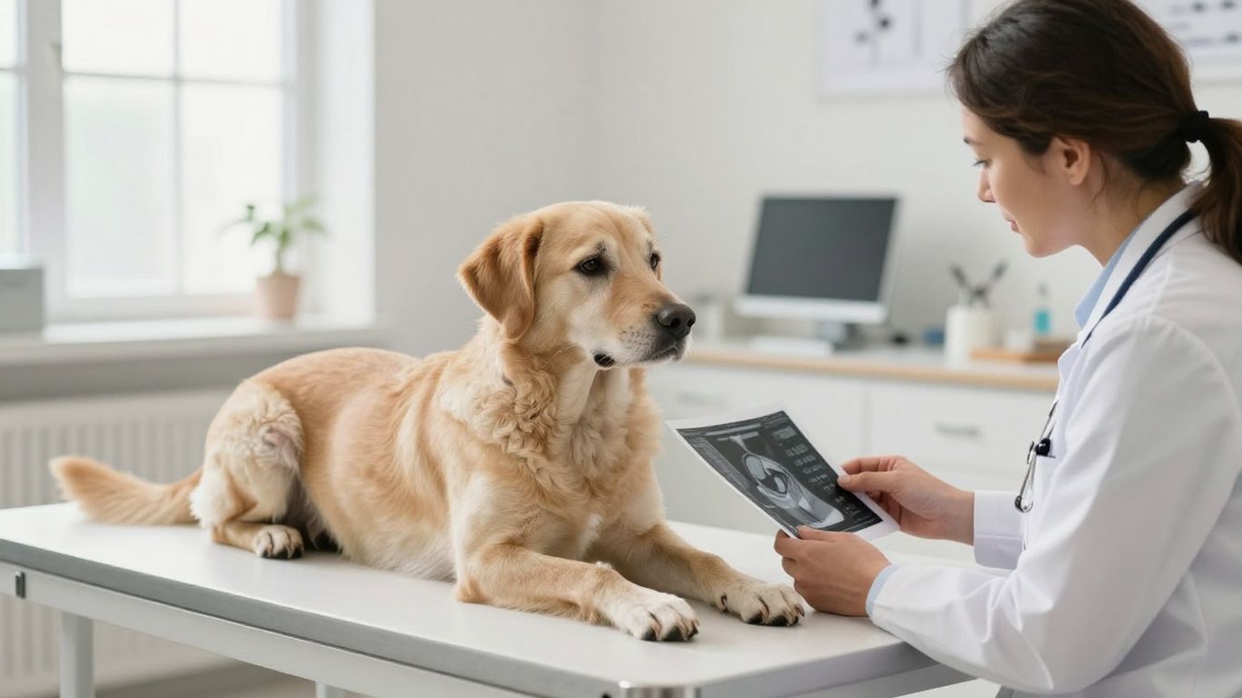
Blood tests may assess general health and help a veterinarian plan sedation, surgery, or other care. Imaging such as radiographs, ultrasound, or advanced scans may be considered when a mass is deep or when there is concern about nearby structures or distant disease.
These tests answer different questions. A normal blood test, for example, does not by itself prove that a localized lump is benign, while an imaging finding may still require tissue confirmation.
Staging and determining whether cancer has spread
If cancer is confirmed or strongly suspected, staging helps estimate its extent. The veterinarian may assess local tissues, regional lymph nodes, and organs elsewhere in the body using an appropriate combination of examination, sampling, blood work, and imaging.
Treatment choices depend on the tumor type, grade, location, spread, and the dog’s overall condition. Ask what each result means before making assumptions based on a single image or report.
What to do after finding a concerning growth
Finding a lump can be frightening, but a calm, organized response is more helpful than repeated checking or home treatment. Make a note of the change, take a few clear photographs, and contact your veterinary practice for guidance. If your dog seems seriously unwell, prioritize immediate care over documentation.
How to photograph and measure the area safely
Photograph the area in natural or even indoor light, using a wider view and a closer view. Include a ruler beside—not pressed against—the growth when possible, and keep the camera level. Ask someone to help steady the dog so you do not restrain or stress them unnecessarily.
Useful records include:
- The date and time of each photograph.
- The growth’s length, width, and approximate height.
- Its body location and whether the surrounding skin looks different.
- Any licking, bleeding, pain, discharge, or change in movement.
Afterward, store the images in date order and share the originals with the veterinary team. Consistency makes progression easier to judge.
Why squeezing or removing a lump at home is risky
Squeezing can cause pain, bleeding, inflammation, or infection, and it may damage cells that a veterinarian needs to evaluate. Cutting or draining a growth at home can also make later treatment more difficult and expose you or your dog to unnecessary risk.
Keep household remedies, essential oils, and human medications away from the area unless specifically advised. Even a soft mass should be handled gently until its identity is known.
Questions to ask your veterinarian
A good appointment leaves you knowing what is known, what remains uncertain, and what happens next. Bring a short list so anxiety does not make you forget the basics. You might ask:
- What are the most likely explanations for this growth?
- Which test would provide the most useful information?
- How soon should testing or treatment occur?
- What changes should make me call sooner?
If you are also organizing family photographs or unrelated visual projects, Karma Hill Photography describes professionally edited images in a private online gallery; that is separate from the clinical records your veterinarian needs, but it illustrates why dated, clearly stored images are easier to review.
Signs that require urgent veterinary care
Seek urgent veterinary help if your dog has trouble breathing, uncontrolled bleeding, severe pain, collapse, marked weakness, repeated vomiting, or cannot eat or drink. A rapidly worsening swelling, a serious wound, or sudden loss of function also warrants prompt advice.
Do not wait for a perfect photograph when your dog’s comfort or safety is at risk. For general household preparation unrelated to veterinary diagnosis, Attic Fanatics discusses scheduling a free assessment; here, the appropriate assessment is a veterinary one.
Conclusion
A photograph can help you notice change, preserve a timeline, and explain a concern, but it cannot tell you whether a dog’s growth is cancerous. Gentle observation and timely veterinary testing are safer than guessing from appearance or treating a lump at home. When in doubt, share the images and your notes with a veterinary professional.
Frequently Asked Questions
Can I identify dog cancer from a photograph?
No. A photograph may show a change in size, color, surface, or location, but it cannot identify the cells involved or determine whether a growth is benign or malignant.
What does a cancerous lump look like on a dog?
It can look like many things, including a firm nodule, a pigmented bump, a nonhealing sore, or a mass beneath the skin. Because harmless conditions can look similar, appearance alone is unreliable.
Should every lump on a dog be tested?
Not every lump requires the same testing, but every new, changing, persistent, or symptomatic lump should be discussed with a veterinarian. The clinician can decide whether monitoring, cell sampling, or biopsy is appropriate.
How quickly should I make a veterinary appointment?
Book a routine appointment soon for a new or changing growth. Seek urgent care for severe pain, uncontrolled bleeding, breathing trouble, collapse, serious weakness, or a sudden loss of function.
Is a soft, movable lump probably harmless?
Soft and movable lumps are often associated with benign conditions, but those features do not prove that a mass is harmless. Proper identification still depends on a veterinary examination and, when indicated, diagnostic sampling.
Can I squeeze a cyst on my dog?
Do not squeeze or drain a suspected cyst at home. The area could become painful or infected, and the procedure could interfere with accurate diagnosis or expose your dog to avoidable complications.
What should I bring to the veterinary appointment?
Bring dated photographs, measurements, the date you first noticed the growth, notes about changes, and a list of medications or supplements. Also describe licking, bleeding, appetite changes, altered activity, or other unusual behavior.



Leave a Reply