Key Takeaways
A new lump can be unsettling, but its appearance cannot tell you whether it is cancer. Careful observation and a timely veterinary examination are the safest next steps.
- Not every lump on a dog is cancerous.
- Fast growth, bleeding, ulceration, pain, or whole-body changes deserve prompt attention.
- Measure and photograph a lump, but do not squeeze, cut, or puncture it.
- A veterinarian may use examination, fine-needle aspiration, biopsy, blood tests, or imaging.
- Treatment depends on the tumor type, grade, stage, and your dog’s quality of life.
Understanding what dog cancer lumps can look like
Dog cancer lumps do not have one consistent appearance. Some are visible on the skin, while others sit beneath it or develop inside the body. A lump may be harmless, but a new or changing growth still deserves a veterinarian’s assessment rather than a guess based on appearance alone.
Common types of lumps found on dogs
Dogs can develop fatty lumps, cysts, warts, inflammatory swellings, benign tumors, and malignant tumors. Lipomas are often soft and movable, while some cysts feel more defined and may contain fluid or thick material. Other growths can feel firm, irregular, attached to deeper tissue, or difficult to see beneath a thick coat.
Age, location, and the dog’s general health can offer useful context, but they do not settle the question. A helpful overview of common dog lumps and cysts can provide background on the range of growths owners may encounter, while a veterinarian determines what a particular lump means.
How cancerous and noncancerous growths may differ
Some cancerous growths enlarge quickly, feel fixed in place, or develop an uneven surface. Others grow slowly and look much like an ordinary benign lump. Conversely, a noncancerous growth can become large, irritated, or troublesome simply because it is located where a collar, harness, or movement repeatedly rubs it.
There is no dependable home rule that separates malignant from benign lumps. Change over time matters, but even a stable lump should be examined if it is new, unusual, or bothersome to the dog.
Why appearance alone cannot confirm a diagnosis
Color, firmness, hair loss, and mobility can help a veterinarian decide what to investigate, but none of them provides a diagnosis by itself. Some tumors change size temporarily, and some harmless growths can look inflamed after being scratched. Trying to identify a tumor from a photograph or description can therefore create either false reassurance or unnecessary fear.
The most useful response is to arrange an appointment and bring a clear record of when the growth appeared. A general guide to when a dog lump may be cancer can help explain why observation is useful but not a substitute for testing.
Where concerning lumps commonly develop
Growths may appear on the trunk, legs, head, neck, paws, or around the mouth. Mammary tissue, the area around the anus, and the skin between the toes also deserve attention during routine checks. Lumps that are hidden under fur or deeper in the body may be noticed only when they cause swelling, discomfort, or a change in behavior.
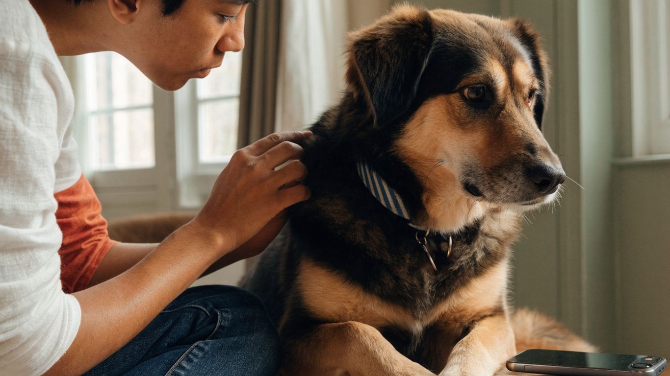
Run your hands gently over your dog during ordinary grooming, and look at areas you may not see from above. Do not assume a lump is unimportant just because it is small or covered by hair.
Recognizing warning signs that need veterinary attention
A lump is more concerning when it changes or begins affecting your dog. That does not prove cancer, but it does raise the value of prompt professional evaluation. If your dog is in severe pain, collapses, has trouble breathing, or has uncontrolled bleeding, contact an emergency veterinary service.

Changes in size, shape, color, or texture
Rapid enlargement is a reason to call the veterinary office rather than waiting for the next routine visit. A lump that becomes harder, develops an irregular outline, turns darker or redder, or seems newly attached to deeper tissue should also be assessed. Changes over days or weeks are especially useful to report.
The absence of change does not guarantee safety. Some tumors remain quiet for a period before becoming more active, and a slow-growing lump can still interfere with movement or require treatment.
Bleeding, oozing, ulceration, or persistent itching
A surface that breaks open, bleeds, leaks fluid, smells unpleasant, or repeatedly scabs needs veterinary attention. Persistent licking, chewing, or scratching may further injure the area and make it harder to judge the original growth. A clean, temporary covering may help prevent self-trauma while you arrange care, but do not apply human medication unless a veterinarian advises it.
Guidance on warning signs in cancer bumps may help you organize what you are seeing. It should not delay a call, particularly when bleeding or rapid change is involved.
Pain, swelling, heat, or reduced movement
Not all cancerous lumps are painful, and pain alone does not identify the cause. Discomfort can arise when a growth presses on tissue, becomes inflamed, ulcerates, or sits near a joint or nerve. Warmth, swelling, limping, reluctance to climb stairs, or a sudden dislike of being touched are meaningful changes to mention.
Avoid repeatedly pressing the area to test it. A dog may react defensively even when the lump itself is not the source of the problem, and handling can worsen irritation.
Whole-body symptoms such as weight loss or fatigue
Reduced appetite, unexplained weight loss, unusual tiredness, vomiting, persistent coughing, or a noticeable change in temperament can accompany many illnesses, including some cancers. These signs are not specific enough to diagnose a tumor, but they make a prompt examination more important. Note when the symptoms began and whether they are getting worse.
A dog that will not eat, seems very weak, or deteriorates quickly should be discussed with a veterinarian the same day. Bring information about medications, appetite, bowel movements, water intake, and any recent changes in the lump.
Evaluating a lump at home without making a diagnosis
Home observation is useful only when it remains gentle and organized. Your role is to document what you see and help the veterinary team understand the timeline. It is not to decide whether the lump is benign or malignant.
How to measure and photograph the growth
Use a ruler to record the lump’s longest dimension and, if possible, its width. Take photographs in consistent lighting with a familiar object or ruler nearby for scale, while keeping the dog comfortable and still. Photographing from the same angle makes gradual changes easier to spot.
Do not shave a thick coat solely to inspect a small bump unless a professional recommends it. The skin may become irritated, and a nervous dog may associate handling with discomfort.
What details to record about changes over time
A short log can prevent uncertain memories from replacing useful observations. Record the date, location, measurements, surface changes, sensitivity, and whether your dog is licking or scratching it. Also note appetite, energy, mobility, and any medications or recent injuries.
These are the most helpful details to bring to the appointment:
- When you first noticed the lump.
- Whether its size or texture has changed.
- Whether it moves under gentle contact or seems fixed.
- Whether the skin is bleeding, open, warm, or irritated.
- Any changes in appetite, energy, weight, or movement.
A simple record gives the veterinarian a clearer starting point and can show whether the growth has changed between visits. Keep the notes factual; words such as “larger” or “more painful” are most useful when paired with dates or measurements.
Why squeezing or attempting to remove a lump is risky
Squeezing can rupture a cyst, spread irritation through surrounding tissue, or cause bleeding. Cutting, draining, tying off, or applying caustic substances can create infection and may complicate later treatment. Some tumors also release substances when disturbed, which can cause a sudden reaction in certain dogs.
Leave sampling and removal to a veterinary professional. Even a lump that looks like a routine cyst may need a different approach.
When multiple lumps may require faster evaluation
Several lumps can be benign, especially in older dogs, but multiple growths should not be dismissed as a single condition. Each may have a different cause. Tell the veterinarian how many you have found, where they are located, and whether they appeared around the same time.
If multiple lumps are accompanied by rapid growth, skin breakdown, swollen lymph nodes, appetite loss, or marked fatigue, call promptly. For broader pet-wellness reading, dog-safe coconut oil guidance is a separate topic and should not be used as a treatment for an unexplained lump.
Understanding the veterinary diagnostic process
The diagnostic process usually begins with a hands-on examination and a careful history. Your veterinarian may recommend sampling the growth even when it appears harmless, because cells are more reliable than appearance. The plan may be simple, or it may involve several tests when the lump is deep, large, fixed, or associated with other symptoms.

What happens during a physical examination
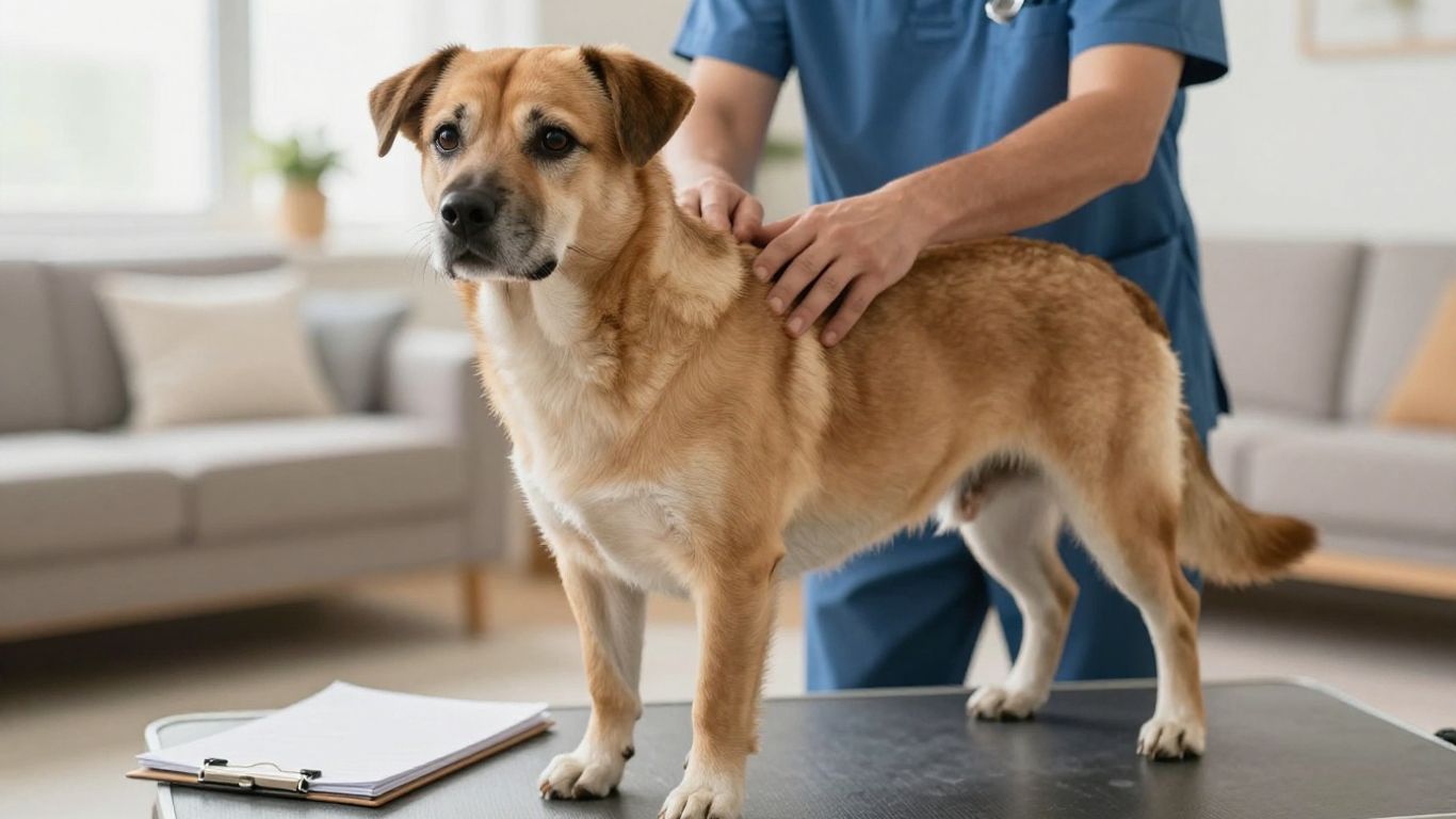
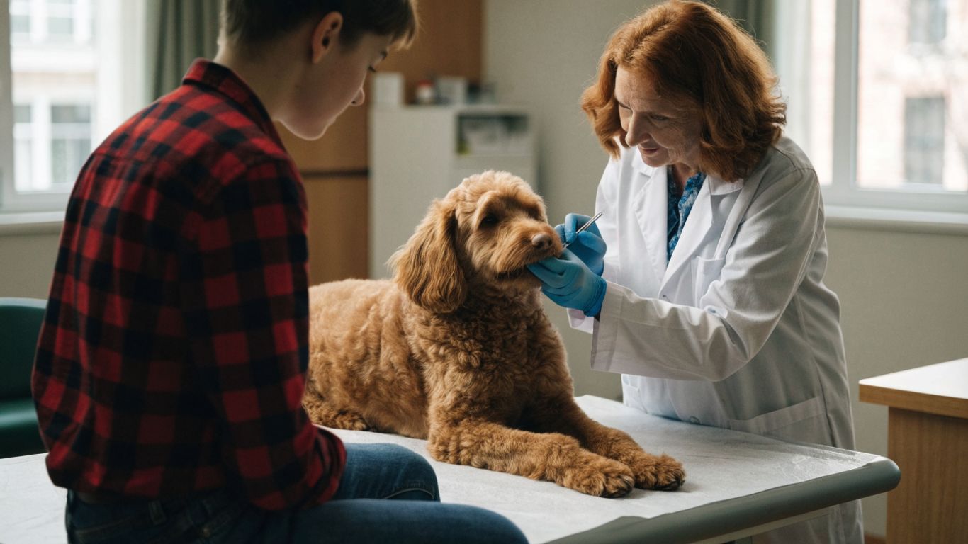
The veterinarian will assess the lump’s size, shape, firmness, mobility, temperature, and relationship to nearby tissue. They may examine the entire dog, including lymph nodes and other skin areas, rather than focusing only on the growth you noticed. Questions about timing, behavior, appetite, and previous medical problems help complete the picture.
Bring your photographs and measurement log. Even small details, such as whether the dog licks the area at night, can influence what the veterinarian recommends next.
How fine-needle aspiration can identify abnormal cells
Fine-needle aspiration uses a small needle to collect cells from the lump for examination. It may help identify certain types of growths, infection, inflammation, or abnormal cells, often without the need for a large procedure. Results are not always definitive, particularly when a sample contains too few cells or does not capture the most representative area.
The veterinarian may recommend repeating the aspiration or moving to a biopsy if the result does not match the lump’s behavior. A reassuring initial sample should still be interpreted in context.
When a biopsy or surgical removal is recommended
A biopsy removes a portion of a growth for laboratory analysis, while excision removes the whole lump when that is appropriate. The choice depends on location, size, suspected tumor type, surgical margins, and whether removing the mass before a diagnosis could make later care more difficult.
Ask whether the tissue will be sent to a veterinary pathologist and when results are expected. A pathology report can describe the tumor type and other features that guide treatment.
The role of blood tests and imaging scans
Blood tests can evaluate general health and help assess whether a dog is ready for anesthesia or treatment. Imaging such as radiographs, ultrasound, CT, or MRI may be considered to understand a mass’s depth, examine nearby structures, or look for disease elsewhere. The right combination depends on the suspected diagnosis and the information needed for a treatment plan.
The tests are not interchangeable. A normal blood panel, for example, does not by itself rule out a localized tumor, while imaging cannot always identify the exact cell type.
Exploring the main types of cancer-related lumps
Cancer-related lumps arise from different tissues and can behave very differently. Some begin in the skin, some in connective tissue, and others affect glands, lymphatic tissue, or internal organs. The names below are categories for discussion, not a way to diagnose a particular dog at home.
Mast cell tumors and their variable appearance
Mast cell tumors are known for having a wide range of appearances. They may resemble a raised button, a soft swelling, or an irritated patch, and their size can fluctuate as nearby cells react. A dog may lick or scratch the area, but some dogs show little outward discomfort.
Because appearance is so variable, sampling is particularly valuable when a veterinarian suspects this type of tumor. A growth that changes size after handling should be reported rather than repeatedly tested at home.
Soft tissue sarcomas beneath or within the skin
Soft tissue sarcomas develop in connective tissues and may form a firm mass under the skin. They can be difficult to see early, especially on a heavily coated dog, and their borders may extend farther beneath the surface than the visible swelling suggests. Growth rate and location help determine how much investigation is needed.
Surgical planning may be important for these masses because the veterinarian may need to consider surrounding tissue. Do not assume that a deep, slow-growing lump is harmless simply because the skin looks normal.
Mammary tumors and changes along the mammary chain
Mammary tumors occur along the line of mammary glands and may be felt as one or more nodules. Check the full mammary chain gently, but avoid excessive manipulation. Any firm nodule, swelling, discharge, or change in the overlying skin should be examined.
The veterinarian may assess nearby lymph nodes and discuss whether sampling or surgery is appropriate. Spaying history, age, overall health, and the number and location of nodules may all affect the workup.
Melanoma, lymphoma, and other less visible cancers
Melanoma may occur in the skin or in areas such as the mouth, where a mass can be harder for an owner to notice. Lymphoma often involves lymph nodes or internal organs rather than presenting as a simple skin lump. Bone and abdominal cancers can also cause swelling, lameness, appetite changes, or other less obvious signs.
A basic guide to canine skin tumors can help explain why different cancers require different tests. If your dog has a new oral swelling, enlarged lymph nodes, persistent lameness, or unexplained illness, schedule an examination even when there is no obvious surface lump.
Knowing what affects treatment decisions
A diagnosis is the beginning of a conversation, not a complete prediction. The veterinary team considers the tumor’s biology, how far it has spread, where it is located, and how the dog is feeling. Your family’s practical limits also belong in that conversation.
How tumor type, grade, and stage guide care
Tumor type describes the cells involved. Grade gives information about how abnormal or aggressive those cells appear under a microscope, while stage describes the extent of disease in the body. Together, these details help estimate likely behavior and identify which treatments may be useful.
Ask the veterinarian to explain unfamiliar pathology terms in plain language. A second pathology review or oncology consultation may be reasonable when the diagnosis is complex or the proposed treatment carries major consequences.
When surgery may be the primary treatment
Surgery may be recommended when a tumor can be removed safely and completely. The surgeon considers the mass’s depth, nearby organs, nerves, blood vessels, and the amount of healthy tissue needed around it. Sometimes a smaller procedure is appropriate; sometimes advanced planning is needed before an operation.
The goal is not simply to make the visible lump disappear. Ask about margins, possible complications, pain control, recovery time, and what will happen if the pathology report shows that additional treatment is needed.
How radiation, chemotherapy, or immunotherapy may be used
Radiation may be considered for local control when surgery cannot remove all affected tissue or when a tumor is in a difficult location. Chemotherapy may be used for cancers that are likely to spread or have already spread, while immunotherapy may be discussed for selected diagnoses. These treatments vary considerably in schedule, side effects, and expected benefit.
A veterinary oncologist can explain whether the aim is cure, long-term control, or comfort. Treatment recommendations should be tied to the specific pathology report rather than to the word “cancer” alone.
Balancing treatment benefits, risks, cost, and quality of life
There is rarely one universally right decision. Consider the expected benefit, travel and scheduling demands, anesthesia risk, side effects, cost, and how treatment may affect the dog’s daily comfort. A plan that preserves appetite, mobility, rest, and companionship may be more appropriate than the most aggressive available option.
Write down the estimates and ask what signs would show that a treatment is helping or harming your dog. Clear expectations can make difficult decisions feel more manageable.
Supporting a dog after a lump is diagnosed
After diagnosis, ordinary care becomes a form of monitoring. Your dog may recover quickly from one procedure and need weeks of support after another. Keep communication open with the primary veterinarian and any oncology team, and ask for written instructions before leaving the clinic.
Preparing questions for the veterinary oncology team
A consultation is easier when your questions are specific. Ask what the diagnosis means, whether the tumor was completely removed, what additional staging is recommended, and what outcome the team considers realistic. You can also ask how quickly a decision is needed and what happens if you choose monitoring or comfort-focused care.
Bring the pathology report, medication list, photographs, and your notes about appetite and behavior. If you are also managing a home project, practical planning resources such as questions for a remodeling contractor are unrelated to veterinary care, but the same habit of comparing written estimates and clarifying responsibilities can be useful when organizing medical costs.
Monitoring the incision or tumor for changes
Check an incision as directed for increasing redness, swelling, discharge, separation, heat, or a bad smell. Prevent licking with the protective method recommended by the clinic, and call if the area looks worse rather than better. If the tumor remains in place, continue recording its size and surface condition without pressing on it.
Photographs can help the veterinary team assess a change between appointments. Do not wait for a scheduled visit if your dog appears painful, the incision opens, or bleeding does not stop.
Managing pain, appetite, activity, and medication
Give medication exactly as prescribed and do not add human pain relievers without veterinary approval. Follow restrictions on running, jumping, stairs, bathing, and wound care. Small, appealing meals may help after anesthesia, but prolonged refusal to eat, repeated vomiting, or marked sleepiness warrants a call.
Keep a simple daily note of medication times, food, water, bowel movements, comfort, and activity. This makes it easier to distinguish expected recovery from a problem that needs attention.
Understanding follow-up visits and recurrence monitoring
Follow-up appointments may include an incision check, physical examination, repeat measurements, blood tests, or imaging. Their timing depends on the diagnosis and treatment, so ask for a written schedule before going home. Even when a tumor has been removed, monitoring helps the team look for recurrence or new growths.
Continue gentle full-body checks after recovery. A new lump is not automatically a recurrence, but it should be documented and reported rather than watched indefinitely.
Conclusion
Dog cancer lumps can look and feel very different, and no home inspection can replace veterinary testing. Notice changes, document them calmly, avoid disturbing the growth, and arrange professional care promptly when warning signs appear. Early, clear communication gives you and your veterinary team the best chance to choose care that fits your dog’s health and your family’s needs.
Frequently Asked Questions
Are all lumps on dogs cancerous?
No. Dogs develop many noncancerous lumps, including fatty growths, cysts, and other benign changes. Because harmless and cancerous growths can look alike, a veterinarian should assess new or changing lumps.
What does a cancerous lump on a dog feel like?
It may feel firm, irregular, fixed, or deep, but it can also be soft or movable. Texture alone cannot identify cancer, so sampling may be needed.
Should a fast-growing lump be treated as urgent?
Rapid growth deserves prompt veterinary attention, especially when it occurs with bleeding, ulceration, pain, appetite loss, weakness, or other sudden changes. Call the clinic for guidance on timing.
Can a dog cancer lump be painful?
Some are painless at first. Pain may develop when a growth becomes inflamed, ulcerated, presses on nearby tissue, or affects movement.
Is it safe to pop or drain a lump at home?
No. Squeezing or puncturing a lump can cause bleeding, infection, irritation, or an inaccurate assessment. Sampling and drainage should be performed by a veterinary professional.
How is a dog lump diagnosed?
A veterinarian may begin with a physical examination and fine-needle aspiration. Depending on the findings, biopsy, surgical removal, blood tests, or imaging may follow.
What should I do if my dog has several lumps?
Record the number, location, size, and changes in each lump, then arrange a veterinary examination. Multiple lumps can have different causes and should be assessed individually.



Leave a Reply